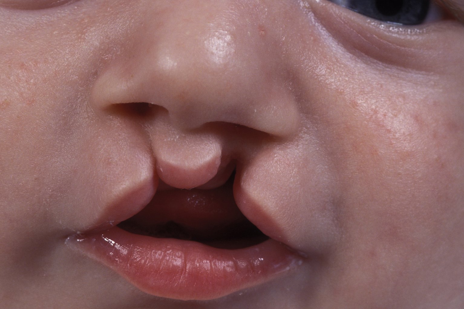
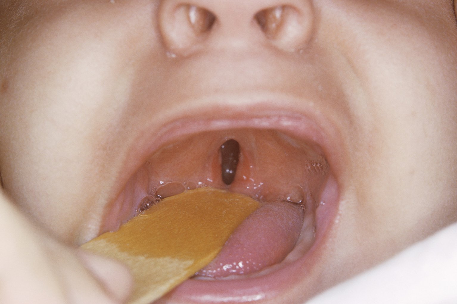
WHAT IS CLEFT LIP AND PALATE?
During first three months of pregnancy there is failure of complete union of the two segment of lip resulting in variable extent of clefting of the Lip.
Cleft palate results in failure of fusion of the two palatine shelves. This failure may be confined to the soft palate alone or involve both hard and soft palate.
INCIDENCE:
Cleft lip and cleft palate is one of the most common congenital anomaly. According to WHO statistics, Pakistan is on fourth number with largest population of cleft lip and cleft palate. Incidence in Pakistan is 1 in 500 live births. 17,000 children are born every day in Pakistan which means 34 children are born daily with Cleft lip or Cleft Palate. That makes 12,400 new clefts just in a year.
CAUSES OF CLEFT LIP AND CLEFT PALATE:
The exact cause or reason for their occurrence is not known. Both Genetic factors and environmental factors are active.
Among the genetic factors cousin marriages is one very important factor Pakistan.
Among the environmental factors, Folic acid deficient is a scientifically proven cause. Others causes are exposure to radiation, high mother’s age, smoking and alcohol consumption, fathers overage, etc.
LUNAR OR SOLAR ECLIPSE has nothing do with clefts. There is no Black magic and there is no association of cleft with exorcism.
RISK OF HAVING A CHILD WITH CLEFT LIP OR CLEFT PALATE:
Sporadic occurrence of cleft is just 2%. If parents have one child with Cleft lip or palate then chances of it having it in second child is 4%. If two children have clefts, then chance s in third child are 9%. If one parent and one child then chances are 14-17%.
CLASSIFICATION:
NORMAL & PATHOLOGICAL ANATOMY OF LIP:
The surface land marks of lip are Cupid’s bow, vermillion, philtrum, philtrum columns, columella, nasal floor and nasolabial angle. The muscle fibers are of orbicularis oris, Levator labia superior is and depressor septi nasi muscle. Blood supply is from superior and inferior labial artery.
The abnormalities in cleft lip are the direct consequence of disruption of the above mentioned muscles of the upper lip and nasolabial region.
Unilateral cleft lip. In the unilateral cleft lip, the nasolabial and bilabial muscle rings are disrupted on one side resulting in an asymmetrical deformity involving the external nasal cartilages, nasal septum and anterior maxilla (premaxilla). These deformities influence the mucocutaneous tissues causing displacement of nasal skin on to the lip and retraction of labial skin, as well as changes to the vermilion and lip mucosa.(Fig.2) In incomplete cleft Simonart’s band consist of a skin bridge across the nasal sill. It does not contain any significant muscle mass. If the cleft is less than two thirds of the lip height, some muscle fibers cross the cleft. (Fig.3)
Bilateral cleft lip. In the bilateral cleft lip, the deformity is more profound but symmetrical. The two superior muscular rings are disrupted on both sides producing a flaring of the nose (due to lack of nasolabial muscle continuity), a protrusive premaxilla and an area of skin in front of the premaxilla, known as the “prolabium”, devoid of muscle. As in the unilateral cleft lip, the muscular, cartilaginous and skeletal deformities influence the mucocutaneous tissues. (Fig. 4)
There are certain associated lip and nasal abnormalities like hypoplastic and flattened alar dome on the affected side, lack of upper lateral cartilage overlap of lower lateral cartilage, subluxed lower lateral cartage with alar base displaced cephalad and posteriorly, hypoplastic bony maxilla, the caudal septum is pulled toward the non cleft side, flattening of the nasal bones and shortened columella especially in bilateral cases.
NORMAL & PATHOLOGICAL ANATOMY OF PALATE:
When the cleft of the hard palate remains attached to the nasal septum and vomer, the cleft is termed incomplete. When the nasal septum and vomer are completely separated from the palatine processes, the cleft palate is termed complete. (Fig.5)
Soft palate. In the normal soft palate, closure of the velopharynx, which is essential for normal speech, is achieved by five different muscles functioning in a complete but coordinated fashion. In general, the muscle fibers of the soft palate are orientated transversely with no significant attachment to the hard palate.
In a cleft of the soft palate, the muscle fibers are orientated in an anteroposterior direction, insetting into the posterior edge of the hard palate
Hard palate. The normal hard palate can be divided into three anatomical and physiological zones .The central palatal flbromucosa is very thin and lies directly below the floor of nose. The maxillary fibromucosa is thick and contains the greater palatine neurovascular bundle. The gingival fibromucosa lies more lateral and adjacent to the teeth.
Sub Mucous Cleft Palate: They are characterized by bifid uvula, notching of the hard palate and zona pellucida. On simple opening of the mouth palate looks normal but patient will be exhibiting hyper nasal speech.
PROBLEMS ASSOCAITED WITH CLEFT LIP AND CLEFT PALATE:
Cosmetic: A child with cleft lip if not repaired at appropriate age suffers from inferiority complex syndrome. He/She shy away from attending school and try to avoid social gathering. They are always victim of jokes and fun form their peers.
Feeding: Most babies born with cleft lip and palate feed well and thrive provided appropriate advice is given and support available. Some mothers are successful in breastfeeding, particularly when the cleft is incomplete and confined to the lip. Good feeding patterns can be established with soft bottles and modified teats. Simple measures, such as enlarging the hole in the teat and feeding at 45º, often suffice. Feeding plates, constructed from a dental impression of the upper jaw, are rarely necessary to improve feeding. Some babies are provided with an active plate which aims not only to improve feeding but also reduce the width of the cleft lip and palate prior to surgery. The long-term benefit of such a regime remains unproven.
Airway: Major respiratory obstruction is uncommon and occurs exclusively in babies with Pierre Robin sequence. Hypoxic episodes during sleep and feeding can be life threatening. Intermittent airway obstruction is more frequent and managed by nursing the baby prone. More severe and persistent airway compromise can be managed by ‘retained nasopharyngeal intubation’ to maintain the airway. Surgical adhesion of the tongue to the lower lip (labioglossopexy) in the first few days after birth is an alternative but less commonly practiced method of management.
Speech: Patients of cleft palate if not operated at correct age, develop velo-pharyngeal in- competency which results in speech that has a hyper-nasality. This is called Rhinolalia.
Ear problems: The muscles which are helpful in closing and opening of Eustachian tube are defective in cleft palate and result in recurrent effusion of middle ear cavity. This might lead to chronic supparative otitis media and subsequently conductive deafness.
Dental: Dental anomalies are common findings in children with cleft lip and/or palate. Various phenomena include delayed tooth development and delayed eruption of teeth; morphological abnormalities are also well documented. The number of teeth may be reduced (hypodontia) or increased (hyperdontia), and occurs most commonly in the region of the cleft alveolus involving the maxillary lateral incisor tooth. These abnormalities can occur in both primary and secondary dentition.
Mid Face Growth: Early palate surgery can adversely affect the growth of maxilla. Therefore palate surgery preferably should be done after 12 months of age.
MANAGEMENT:
Management is multi-disciplinary and involves Pediatrician, Orthodontist, Plastic Surgeons, Maxillofacial surgeon, Dental surgeon, Psychiatrist, Social worker and Speech therapist.
Management starts immediately after birth and continue till the child enters adult hood.
Immediately after birth the role of Pediatrician and Orthodontist starts. Pediatrician reassures the parents and makes sure that child gets proper nutrition and grows normal weight. Orthodontist role starts in doing naso alveolar molding so that different maxillary segments are brought in alignment and shape of the nose is not distorted.
Sometimes help of Otolarngologist is seeked to improve Eustachian function.
When the child is of three months of age lip repair is done. When child is of 9 months to 18 moths of age then palate repair is done. Then the child is observed for growth of the face and speech. If any problem in lip or nasal growth occurs, then lip nose revision is done between 6 to years of age. If speech problem, because of velo-pharyngeal in- competency is encountered then pharyngoplasty is done after six years of age.
At the age of mixed dentition alveolar bone grafting is done. At puberty definitive open Rhinoplasty is done and sometimes, if need is there Orthognathic surgery is indicated.
Meticulous record-keeping of photography, radiology, dental casts and speech recording are indispensable, and permit regular audit and improved outcomes.
PRINCIPLES OF SURGCILA REPAIR:
1.OF CLEFT LIP:
i) Excision of tissue should be as little as possible.
ii) Cupid’s bow should be reconstituted.
iii) Natural landmarks should be preserved and correctly positioned.
iv) Scaring should be minimal.
v) The lip must be sutured in three layers.
vi) Always restore functional continuity of the muscles.
2.OF CLEFT PALATE:
i)The abnormal attachments of levator veli palatini and tensor veli palatini should be detached from posterior border of hard palate and brought in midline.
ii) The palate should be closed in three layers i.e. nasal, muscle and buccal layers.
iii) Minimal raw areas of bare palatal bone should be left.
PRINCIPLES OF SURGCILA REPAIR:
Fall into four types:
- Rose-Thompson repair: involves simple pairing and suturing.
- Tennison Randal: involves inter positioning of local triangular flaps.
- LeMesurier& Hagedorn: involves inter-positioning of quadrangular flaps.
- Millard: This method of rotation and advancement is the most popular and most practiced method. Its modifications by Noordhoof and Muller are also very commonly employed. This method involves rotation of medial lip downward to fill the cleft defect. A small pennant- shaped “C” flap can either be rotated to create the nasal sill or used to lengthen the columella. Muscular continuity is achieved by subperiosteal undermining over the anterior maxilla. Nasolabial muscles are anchored to the premaxilla with non-resorbable sutures. Oblique muscles of orbicularis oris are sutured to the base of the anterior nasal spine and cartilaginous nasal septum. Closure of the cleft lip is completed by suturing the horizontal fibers of orbicularis oris to achieve a functioning oral sphincter.
Advantages are that it is simple, easy to perform ,does not entail complicated measurements, does not violate cupid’s bow or philtral column, leaves inconspicuous scar and easily revisable.
Disadvantages are inadequate flap rotation leading to notching and inadequate vertical lip length. It is difficult for wider clefts. (Fig.6)
Bilateral cleft lip Repair: are more difficult to repair as Prolabium is often protruding. This can be brought in alignment by pre surgical orthopedic treatment or by lip adhesion or by simple tapping of the prolabium.
During surgery prolabium skin is used to remake philturm. Prolabial vermillion issued to reconstruct the labial sulcus. The final lip vermillion is composed only of vermillion from the lateral lip segments, not from the prolabium.
PROCEDURES FOR THE CLEFT PALATE SURGERY
Cleft palate closure can be achieved by one- or two-stage palatoplasty. The surgical principle is mobilization and reconstruction of the aberrant soft palate musculature, together with closure of the residual hard palate cleft by minimal dissection and subsequent scar formation. Bilateral bipedicled mucoperiosteal flaps are raised on greater palatine vessels, brought in midline and sutured in three layers after receiving lateral relaxing incisions bilaterally. (Fig.7)
Excess scar formation in the palate adversely affects growth and development of the maxilla. The philosophy of two-stage closure encourages a physiological narrowing of the hard palate cleft to minimize surgical dissection at the time of the second procedure.
The most commonly employed technique is Von Langenbeck followed by Veau Wardill technique. Furlow double opposing Z palatoplasty is meant for posterior soft cleft palate only.
CLEFT ALVEOLUS SURGERY:
Mucoperiosteal flaps are raised and inset as advancement flaps. Bone grafting is performed.
COMPLICATIONS OF CLEFT PALATE SURGERY:
Bleeding, Respiratory obstruction, infection dehiscence and oro-nasal fistula formation. Mid face growth problems and velo pharyngeal incompetence can also occur.
Velo pharyngeal insufficiency: (VPI) is the incomplete closure of the velum against the posterior pharyngeal wall. It is found in 20% of the patients operated for cleft palate surgery. Its is corrected by surgery of pharyngeal flaps and sphincter pharyngoplasty


